Call for Paper Track: 16 Electron Microscopy
12th Emirates Pathology & Digital Pathology Conference on December 21-23, 2022 in Dubai, UAE
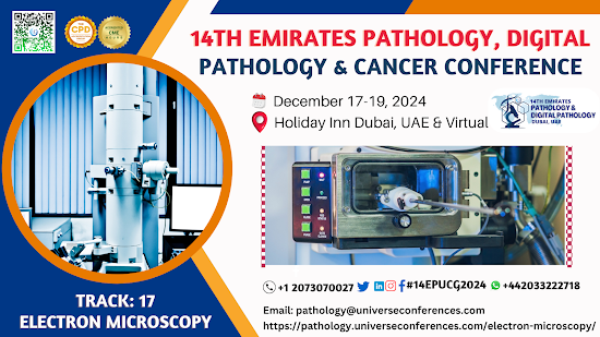
What is Electron Microscopy?
High resolution photographs of both biological and
non-biological specimens can be obtained using electron microscopy (EM). It is
used in biomedical research to examine the precise structure of tissues, cells,
organelles, and macromolecular complexes. The utilisation of electrons—which
have extremely short wavelengths—as the source of illuminating light
contributes to the great resolution of EM pictures. To address particular
issues, electron microscopy is combined with a range of auxiliary procedures
(such as thin sectioning, immuno-labeling, and negative staining). The
structural underpinnings of cell activity and illness are crucially revealed by
EM imaging.
The transmission electron microscope (TEM) and the scanning
electron microscope are the two primary varieties (SEM). Thin specimens (such
as molecules, tissue slices, etc.) that allow electrons to pass through and
produce a picture are observed using a transmission electron microscope. The
ordinary (compound) light microscope and the TEM are both comparable in many
ways. TEM is used, among other things, to visualise the interior of cells in
thin sections, the structure of protein molecules in contrast to metal
shadowing, the arrangement of molecules in viruses and cytoskeletal filaments
in preparation for negative staining, and the positioning of protein molecules
in cell membranes (by freeze-fracture).
.png)
12th Emirates Pathology & Digital Pathology Congress
on December 21-23, 2022 in Dubai, UAE
The emission of secondary electrons from a specimen's
surface is a prerequisite for conventional scanning electron microscopy. A
scanning electron microscope is the equivalent of a stereo light microscope in
the EM because of its superior depth of focus. It offers intricate pictures of
the surfaces of cells and entire organisms, which TEM cannot do. Additionally,
it can be utilised for process control, particle size analysis, and counting.
Because the image is created by rastering a focussed electron beam across the
specimen's surface, it is known as a scanning electron microscope. At each
point in the raster, particles (such as low energy particles) are emitted as a
result of the principal electron beam's interaction with atoms close to the
surface.
These can be gathered using a variety of detectors, and the
brightness at each analogous position on a cathode ray tube can be calculated
from their relative number. The final image is an enlarged image of the
specimen since the size of the raster at the specimen is significantly smaller
than the viewing screen of the CRT. SEMs that are properly set up (with
secondary, backscatter, and X-ray detectors) can be used to examine specimen
topography, atomic composition, as well as, for instance, the distribution of
immuno-labels on the specimen's surface.
12th Emirates Pathology & Digital Pathology Event
on December 21-23, 2022 in Dubai, UAE
Benefits of using an electron microscope
The fundamental benefits of electron microscopy are
numerous. These consist of:
Magnification and
improved resolution are possible thanks to the use of electrons rather than
light waves, allowing for the analysis of previously invisible structures.
Images taken using an electron microscope have a resolution of up to 0.2 nm,
making them 1000 times more detailed than those taken with a light microscope.
Numerous uses -
Electron microscopy is used in a wide variety of study domains, including
technology, industry, biomedical science, and chemistry. Examples of
applications include semiconductor inspection, the production of computer
chips, quality assurance and control, and atomic structure analysis structures,
and drug development
High-quality photos:
With the right training, an operator of an electron microscope can use the
system to create extremely detailed photographs of structures that are of a
high quality, exposing intricate and delicate structures that other methods
might find difficult to replicate.
12th Emirates Pathology & Digital Pathology Summit
on December 21-23, 2022 in Dubai, UAE
Different Electron Microscope Types
The transmission electron microscope (TEM), scanning
electron microscope (SEM), and reflection electron microscope are only a few
examples of the various types of electron microscopes (REM.) In this article,
each of these varieties of the electron microscope will be detailed in further
detail.
Transmission
electron microscope (TEM)
The first form of electron microscope was the transmission
electron microscope, which uses a high voltage electron beam to illuminate the
object.
The electron beam is created by an electron gun. The electron
beam's source, a tungsten filament cathode, is often mounted on the gun. The
electron beam is focused with the use of electrostatic and electromagnetic
lenses and is accelerated by an anode.
the spatial variation can be investigated. Placing a
photographic film into an electron beam to capture the image is yet another way
to capture the image. The image can also be seen on a computer screen in real
time using a digital camera.
Historically, transmission electron microscope resolution
has been constrained by spherical aberration. However, recent advancements have
made it possible to get around this problem and boost resolution via hardware
spherical aberration correction. As a result, it is now possible to create
images with resolutions lower than 0.5 angstroms and magnifications greater
than 50 million times.

12th Emirates Pathology & Digital Pathology Exhibition
on December 21-23, 2022 in Dubai, UAE
Scanning
electron microscope (SEM)
Raster scanning was a method used by the scanning electron
microscope to create enlarged pictures of the sample. It focuses an electron
beam that travels across the specimen's rectangular region, losing energy as it
does so. Heat, light, secondary electrons, backscattered electrons, and other
types of energy are produced from the energy.
However, it is advantageous because it makes use of surface
processes, which enables it to produce images of huge samples with a wider
depth of field and a maximum size of several centimetres. As a result, the
images produced by a SEM may be accurate depictions of the specimen's true
shape.
Reflection
electron microscope (REM)
A beam of elastically dispersed electrons that is reflected
off of the item under examination is detected using a reflection electron
microscope. This kind of microscopy frequently employs the reflection
high-energy electron diffraction (RHEED) and reflection high-energy loss
spectroscopy (RHELS) techniques.
12th Emirates Pathology & Digital Pathology Seminar on December
21-23, 2022 in Dubai, UAE
Important Information:
Conference Name: 12th Emirates Pathology &
Digital Pathology Utilitarian Conference
Short
Name: 12EPUCG2022
Dates December
21-23, 2022
Venue: Dubai, UAE
Scientific Program: It will only
include plenary speakers, keynote speakers, panel discussions and presentations
in parallel sessions.
Audience: Global Leaders,
Industrialists, Business Delegates, Students, Entrepreneurs, Executives
Email: pathology@universeconferences.com
Visit: https://pathology.universeconferences.com/
Pathology Experts:https://pathology.universeconferences.com/pathology-experts/
Call for Papers: https://pathology.universeconferences.com/submit-abstract/
Register here: https://pathology.universeconferences.com/registration/
Call Us: +12073070027
WhatsApp
Us: +4420332227110
Pathology Medical Conferences, Histopathology
meeting, Immunology
conference, clinical
Pathology webinars, Plant
Pathology Conferences, CME
Pathology Conferences, Pathology
Congresses, World
Pathology Congress, Clinical
Pathology, Laboratory
Medicine Conference, Digital Pathology exhibition
Reference pathology and digital pathology
UCGconferences press releases and blogs
Medium: https://medium.com/@traviis.stork/call-for-paper-track-3-breast-pathology-837328eaad1
Globa lhealth : https://globalhealthtrainingcentre.tghn.org/community/blogs/your_posts/
Linked In: https://www.linkedin.com/pulse/call-paper-track-3-breast-pathology-dr-travis-stork-1f
Tumbler: https://www.tumblr.com/blog/view/digitalpathologyucgconferences/689005131446059008?source=share


Comments
Post a Comment