Posts

Showing posts from October, 2023

The Role of Pathologists in Diagnosing Disease: Deadline Alert! Submit an abstract at the 13EPUCG2023 Conference from Dec 15–17, 2023, in Dubai & Virtual.
Image
 Pathologists are often the unsung heroes in the field of medicine, playing a critical role in diagnosing diseases and guiding patient care. In this blog post, we will shed light on the essential functions of pathologists in the diagnostic process and their impact on patient outcomes. ·         Defining Pathology : Start by explaining what pathology is and the primary responsibilities of pathologists. Highlight their expertise in understanding the mechanisms of disease. ·         Histopathology and Microscopic Examination : Discuss how pathologists use microscopes to examine tissue samples, identifying abnormalities, such as cancer, infections, and inflammatory conditions. ·         Cytopathology and Fine Needle Aspiration ( FNA ) : Explain how pathologists evaluate individual cells and use techniques like FNA to diagnose conditions like thyroid nodules and breast lumps. ·         Autopsy and Post-Mortem Examination : Explore the role of pathologists in performing autopsies to
Pathology Lab Techniques: From Microscopes to Genetic Testin

Image
  In the realm of pathology, the laboratory is where the magic happens. Pathologists and lab technicians use a variety of techniques and tools to uncover the mysteries of disease. In this blog post, we'll take a journey through the various lab techniques that play a pivotal role in pathology : Microscopy inPathology : Begin with an exploration of how traditional light microscopy remains the backbone of pathology, allowing pathologists to examine tissue and cellular structures in detail. Histopathology : Delve into the art of preparing and staining tissue samples to visualize cellular structures and identify abnormalities, making it the cornerstone of diagnosing diseases. Cytopathology : Discover how the examination of individual cells from various bodily fluids, like Pap smears, plays a crucial role in cancer detection and diagnosis. Immunohistochemistry (IHC): Learn how IHC uses antibodies to identify specific proteins in tissue samples, aiding in the diagnosis of various
Image
Pathology is a field that continuously evolves with advancements in technology and scientific understanding. In this blog post, we'll explore the exciting emerging trends and technologies that are shaping the future of pathology:  Digital Pathology : The transition from traditional microscopy to digital pathology is changing how pathologists work. Explore the benefits of digital slides, AI-assisted analysis, and remote consultations.  Artificial Intelligence (AI) in Pathology: AI is poised to revolutionize pathology by assisting in diagnosing diseases, identifying patterns, and improving the accuracy and speed of analysis. Learn about the latest AI applications in pathology.  Liquid Biopsies: Liquid biopsies involve testing blood or other bodily fluids to detect diseases, including cancer. Discover how this non-invasive method is transforming cancer diagnosis and monitoring.  Molecular Pathology: Genomic and molecular techniques are providing unprecedented insights into

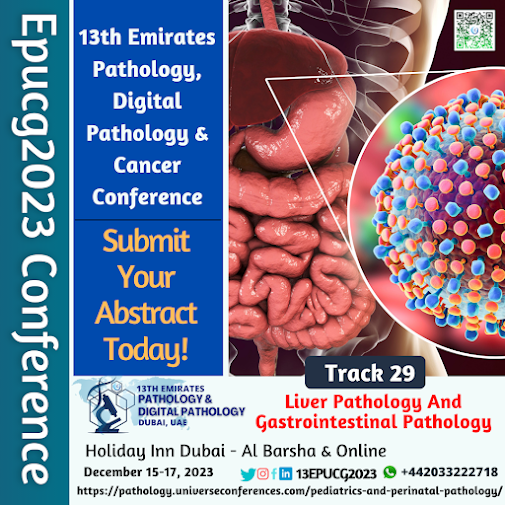
ما هي أمراض الكبد وأمراض الجهاز الهضمي

Image
أمراض الكبد وأمراض الجهاز الهضمي هي مجموعة واسعة من الحالات والاضطرابات التي تؤثر على الكبد والجهاز الهضمي. إليك بعض الأمثلة على أمراض كل منهما: أمراض الكبد: • التهاب الكبد الفيروسي: يشمل الفيروسات مثل فيروس التهاب الكبد B والتهاب الكبد C والتهاب الكبد A، ويمكن أن يتسبب في التهاب الكبد وتلفه. • تليّف الكبد (Cirrhosis): حالة تتسم بتلف نسيج الكبد واستبداله بنسيج ندبي، وغالبًا ما تكون نتيجة لالتهاب مزمن أو استهلاك الكحول بشكل مفرط. • الدهون في الكبد (Fatty Liver Disease): تتمثل في تراكم الدهون في خلايا الكبد، وهناك اختلاف بين الدهون الكبدية غير الكحولية والدهون الكبدية الكحولية. • ورم الكبد (Liver Tumor): يمكن أن يكون الأورام الكبدية سرطانية (سرطان الكبد) أو حميدة (ورم الكبد الحميد). أمراض الجهاز الهضمي: •  القرحة المعديّة والاثنى عشريّة (Peptic Ulcers): قرحة تظهر عادة في المعدة أو الأمعاء الدقيقة نتيجة التآكل البطئ للأغشية المخاطية. •  التهاب الأمعاء (Inflammatory Bowel Disease - IBD): يشمل مرضي كرون والتهاب القولون التقرحي وهما حالتين مزمنتين تصيب الأمعاء وتسبب التهابًا وقرحًا.

What is Pediatric and perinatal pathology curriculum to know more Attend_#13EPUCG from December 15–17, 2023, in Dubai, UAE, & Virtual.

Image
 As of my last knowledge update in September 2021, the curriculum for pediatric and perinatal pathology typically encompasses the study of diseases and conditions that affect fetuses, neonates, infants, children, and adolescents. This is a specialized field of pathology that focuses on the diagnosis and understanding of diseases and abnormalities in these age groups.  While the specific curriculum can vary by institution, some common topics and areas of study in pediatric and perinatal pathology include: 1.    Fetal Pathology: This involves the study of diseases, abnormalities, and conditions that affect fetuses during pregnancy. This can include genetic disorders, structural anomalies, and developmental abnormalities. 2.    Perinatal Pathology: This area covers diseases and conditions that occur around the time of birth, including stillbirths, neonatal deaths, and complications during delivery. 3.    Pediatric Neoplasms: The study of childhood cancers, their causes, classification,

نظرة عامة على ماهية أمراض الدم وأهميتها في تشخيص وإدارة اضطرابات الدم.

Image
  نظرة عامة على ماهية أمراض الدم وأهميتها في تشخيص وإدارة اضطرابات الدم. نظرة عامة على أمراض الدم وأهميتها في تشخيص وإدارة اضطرابات الدم: أمراض الدم هي مجموعة متنوعة من الاضطرابات التي تؤثر على مكونات الدم، وتشمل الأحمر، والأبيض، والصفائح، والعوامل المتعلقة بالتخثر. هذه الاضطرابات يمكن أن تكون وراثية أو مكتسبة، حادة أو مزمنة، وقد تؤدي إلى مشاكل صحية خطيرة إذا لم يتم التعرف عليها وعلاجها بشكل صحيح. لهذا السبب، فإن فهم أمراض الدم وأهميتها في التشخيص والإدارة يعتبر أمرًا بالغ الأهمية. أهمية أمراض الدم في التشخيص والإدارة: تشخيص دقيق: أمراض الدم تتطلب تقنيات تشخيص دقيقة وخبرة متخصصة. تحديد النوع والسبب الكامن للمشكلة هو الخطوة الأولى في تقديم العلاج الصحيح. علاج موجه: تعتمد استراتيجيات العلاج على نوع وخصائص الاضطراب الدموي. بعض الأمراض تتطلب متابعة دورية وإدارة أمراض مزمنة، بينما يمكن أن تتطلب الحالات الحادة تدخلاً فوريًا مثل التحول النقوي الحاد. التقدم في العلاج: تم تطوير العديد من العلاجات المبتكرة لأمراض الدم في السنوات الأخيرة، بما في ذلك العلاج الجيني وعلاجات العقاقير الهدف

What is SEM vs TEM electron microscopy?

Image
  What is SEM vs TEM electron microscopy? Scanning Electron Microscopy (SEM) and Transmission Electron Microscopy (TEM) are both powerful techniques used in electron microscopy to visualize the microstructure and nanoscale details of various materials. However, they differ in their fundamental principles, applications, and how they produce images. Scanning Electron Microscopy (SEM): Principle: SEM works by scanning a focused electron beam across the surface of a specimen. When the electrons interact with the sample, various signals are generated, including secondary electrons, backscattered electrons, and characteristic X-rays. These signals are detected and used to create an image. Depth of Imaging : SEM provides 3D-like images of the surface morphology of a sample. It is particularly useful for examining the external features and topography of specimens. Sample Preparation: Samples for SEM need to be coated with a thin layer of conductive material (e.g., gold or carbon)