What is Electron Microscopy
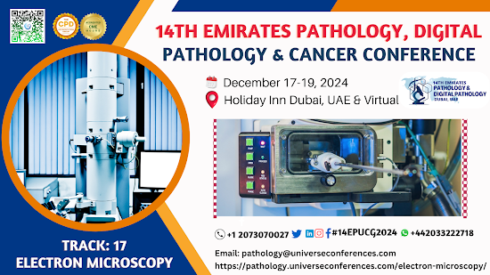
Electron microscopy is a powerful technique used to study the ultrastructure of cells and tissues. It uses a beam of electrons to create00 high-resolution images, allowing for detailed examination of cellular organelles, membranes, and other structures at the nanometer scale. There are two main types of electron microscopy: transmission electron microscopy (TEM) and scanning electron microscopy (SEM). TEM is used to visualize internal structures of cells and tissues, while SEM provides detailed 3D surface images. Both techniques are widely used in various fields of research, including cell biology, pathology, materials science, and nanotechnology.
Electron microscopy is a technique that uses a beam of accelerated electrons to examine objects on a very fine scale. It allows for much higher magnification and resolution compared to light microscopy, enabling the visualization of very small structures such as individual cells, organelles, and even large molecules. There are two main types of electron microscopy:
Transmission
Electron Microscopy (TEM): In TEM, electrons pass through a thin specimen, and the resulting image
is formed by the interaction of electrons transmitted through the specimen.
This technique provides detailed information about the internal structure of
the specimen and is commonly used in biological and materials science research.
Scanning
Electron Microscopy (SEM): SEM scans a focused beam of electrons across the surface of a specimen,
and the interactions of these electrons with the sample surface generate
signals that can be used to create an image. SEM is particularly useful for
studying the surface morphology of specimens in great detail.
Electron microscopy has been invaluable in advancing our understanding of
biological structures, materials science, and nanotechnology, among other
fields, due to its ability to provide high-resolution images at the nanometer
scale.
Conference Name: 14th Emirates
Pathology, Digital Pathology & Cancer Conference
Short Name: 14EPUCG2024
Dates: December 17-19, 2024
Venue: Holiday Inn Dubai, UAE &
Online
Email: pathology@universeconferences.com
Visit: https://pathology.universeconferences.com/
Submit here: https://pathology.universeconferences.com/submit-abstract/
Register here: https://pathology.universeconferences.com/registration/
Online Registration here:https://pathology.universeconferences.com/virtual-registration/
Call Us: +12073070027
WhatsApp us athttps://wa.me/442033222718?text=
#ElectronMicroscopy #TEMmicroscopy
#SEMmicroscopy #ElectronMicrograph #Ultrastructure #Nanotechnology
#MicroscopyArt #MicroscopyTechniques #CellularUltrastructure
#HighResolutionImaging


.png)
Comments
Post a Comment