What is SEM vs TEM electron microscopy?
What is SEM vs TEM electron microscopy?
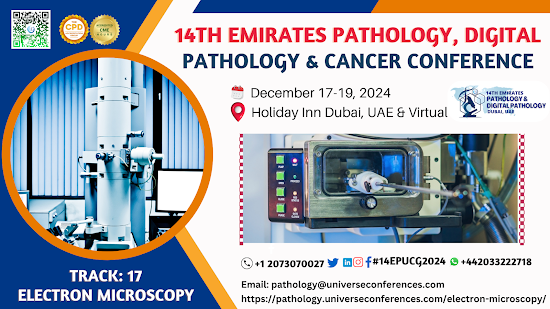
Scanning Electron Microscopy (SEM) and Transmission Electron
Microscopy (TEM) are both powerful techniques used in electron microscopy to
visualize the microstructure and nanoscale details of various materials. However,
they differ in their fundamental principles, applications, and how they produce
images.

Scanning Electron Microscopy
(SEM):
Principle: SEM
works by scanning a focused electron beam across the surface of a specimen.
When the electrons interact with the sample, various signals are generated,
including secondary electrons, backscattered electrons, and characteristic
X-rays. These signals are detected and used to create an image.
Depth of Imaging:
SEM provides 3D-like images of the surface morphology of a sample. It is
particularly useful for examining the external features and topography of
specimens.
Sample Preparation:
Samples for SEM need to be coated with a thin layer of conductive material
(e.g., gold or carbon) to enhance image quality and prevent charging artifacts.
They also need to be dry and non-conductive materials may require sputter
coating.
Resolution:
SEM can achieve resolutions in the nanometer range (typically 1-10 nm),
depending on the instrument and sample.
Applications:
SEM is often used in fields such as materials science, biology, geology, and
forensics to study surface structures, particles, and fractured surfaces.
Transmission Electron
Microscopy (TEM):
Principle:
TEM transmits a beam of electrons through an ultrathin specimen (on the order
of tens of nanometers thick). The electrons that pass through the specimen
interact with it, and the transmitted electrons are used to form an image.
Depth of Imaging:
TEM provides 2D images of the internal structure of a specimen. It allows for
detailed examination of the ultrastructure, including cellular organelles,
crystal lattices, and nanoscale features.
Sample Preparation:
TEM samples require extremely thin sections, typically achieved using
techniques like ultramicrotomy for biological specimens or focused ion beam
(FIB) milling for solid materials. The specimens must be electron-transparent.
Resolution: TEM
offers extremely high resolution, often down to the sub-angstrom level (0.1
nm), making it suitable for studying individual atoms.
Applications:
TEM is widely used in materials science, biology, nanotechnology, and materials
characterization to examine the internal structure of materials, nanoparticles,
biological cells, and viruses.
In summary, SEM is primarily used for surface imaging and
provides 3D-like information, while TEM is used for detailed internal imaging
and provides very high-resolution 2D images. The choice between SEM and TEM
depends on the specific research or analysis requirements and the nature of the
sample being studied.
----
Greetings. We are organizing an in-person CME/CPD accredited The 13th
Emirates Pathology, Digital Pathology & Cancer Conference, Which is
scheduled to from December 15-17, 2023, in Dubai UAE & Online. And we
invite you to attend as a speaker/listener/poster presenter/Exhibitor.
Please let me know if you are interested.
If you have any questions, please Contact us.
Email:
pathology@universeconferences.com
WhatsApp: https://wa.me/442033222718?text=
Visit our Website Here: https://pathology.universeconferences.com/
MedicalIndustry PathologyExhibitor PathologyConference ExhibitHall TradeShow PathologyProducts ExhibitorBooth PathologyTechnology PathologySolutions MedicalExhibitor PathologyInnovation ExhibitionStand MedicalDevices PathologyEquipment ExhibitorShowcase PathologyDemonstration PathologyExpo ExhibitorMarketing PathologyIndustry ExhibitionNetworking PathologyExhibitorLife
PathologyDirector PathologyLeadership PathologyManagement PathologyDepartment PathologyAdministration PathologyDirectorRole PathologyCollaboration PathologyExcellencePathologyCareer
Pathologyconference Emiratespathologyconference Conference2023 pathologyconference2024
PathologyInnovation PathologySpecialists MedicalDiagnostics HealthcareSolutions PathologyConsulting PathologyExpertise PathologyIndustry MedicalResearch PathologyLeadership PathologyCare PathologyQuality


Comments
Post a Comment