Posts

Understanding Different Types of Cancer: A Comprehensive Guide

Image
Cancer is a broad term for a group of diseases characterized by the uncontrolled growth and spread of abnormal cells. There are over 100 different types of cancer, each classified by the type of cell initially affected. Understanding the various types of cancer can help in recognizing symptoms, seeking appropriate treatment, and fostering awareness. Here’s an overview of some of the most common and notable types of cancer: 1. Breast Cancer Overview: Breast cancer develops in the cells of the breasts, most commonly in the ducts or lobules. It primarily affects women but can also occur in men. Symptoms: A lump in the breast or underarm Changes in breast shape or size Nipple discharge Skin changes on the breast Risk Factors: Genetics, age, hormonal factors, lifestyle choices Screening: Mammograms, breast self-exams, clinical breast exams 2. Lung Cancer Overview: Lung cancer begins in the lungs and is often associated with smoking, although non-smokers can also develop it. T

New Technologies in Pathology: Highlights from the Pathology, Digital Pathology & Cancer Summit

Image
1. AI-Powered Diagnostic Tools: At the forefront of the Digital Pathology Summit were AI-powered diagnostic tools. These technologies are revolutionizing pathology by providing enhanced image analysis, pattern recognition, and predictive analytics. AI algorithms can now assist pathologists in identifying abnormalities with greater accuracy and speed, reducing diagnostic errors and improving patient outcomes. 2.  Whole Slide Imaging (WSI): Whole Slide Imaging was a hot topic, showcasing how it enables the digitization of entire pathology slides. This technology allows for easy storage, sharing, and analysis of digital slides, facilitating remote consultations and second opinions. WSI also integrates seamlessly with AI tools, further enhancing diagnostic capabilities. 3.  Digital Pathology Workflow Solutions: Innovations in digital pathology workflows were highlighted, focusing on streamlining the entire process from slide scanning to diagnosis. These solutions aim to improve efficiency,

"Ensuring Medication Safety: Best Practices for Patients and Caregivers"

Image
  Medication safety is a critical aspect of healthcare that often doesn't receive the attention it deserves. While medications are essential for treating various health conditions, improper use can lead to serious health risks, including adverse drug reactions, hospitalizations, and even fatalities. Here, we’ll explore key strategies to ensure medication safety for both patients and caregivers. Use a Medication Schedule: Keeping a medication schedule can help ensure you take your medications at the right times. Tools like pill organizers, apps, or alarm reminders can be very effective. Store Medications Properly: Store medications in a cool, dry place, away from direct sunlight. Ensure they are out of reach of children and pets. Some medications may require refrigeration—check the storage instructions. Dispose of Medications Safely: Do not flush medications down the toilet unless the label specifically instructs you to do so. Utilize local medication take-back programs or fo

Speculate on the future of immunopathology research, including potential breakthroughs and areas of growth

Image
Immunopathology is a branch of pathology that deals with the study of the immune system in health and disease. It focuses on understanding the mechanisms by which the immune system responds to various stimuli, such as infectious agents, toxins, or self-antigens, and how these responses can lead to disease. In immunopathology, researchers and healthcare professionals study the structure and function of the immune system, including its cells, molecules, and organs, such as the thymus, spleen, and lymph nodes. They also investigate how the immune system interacts with other systems in the body, such as the endocrine and nervous systems. One of the key aspects of immunopathology is the study of immunological disorders , including autoimmune diseases, hypersensitivity reactions, immunodeficiencies, and transplant rejection. By understanding the underlying immunopathological mechanisms, researchers can develop better diagnostic tools and treatments for these conditions. Here are some spec

#Callforstudent

Image
 Early Bird Deadline: May 15th, 2024 #Callforstudent Grab your Speaker/Listener/Poster slot for CME/CPD accredited 14th Emirates Pathology, Digital Pathology & Cancer Conference from December 17-19, 2024, at Holiday Inn Dubai, UAE & Virtual. Register Now: https://pathology.universeconferences.com/special-conference-registration/ WhatsApp us: https://wa.me/442033222718?text= #histopathology #Medicalstudent #Dubaimedicalstudent #Medicalstudent #pathology #medicine #cytopathology #pathologist #histopathology #histology #EPUCG #Medicalstudent #Studentpath

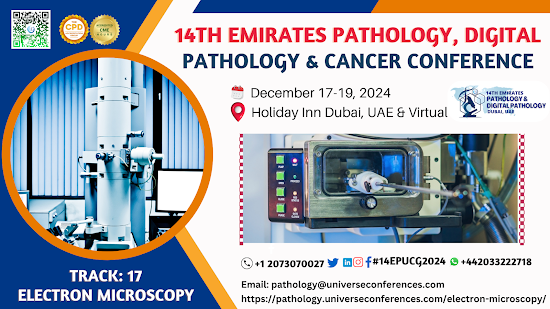
What is Electron Microscopy

Image
Electron microscopy is a powerful technique used to study the ultrastructure of cells and tissues. It uses a beam of electrons to create00 high-resolution images, allowing for detailed examination of cellular organelles, membranes, and other structures at the nanometer scale. There are two main types of electron microscopy: transmission electron microscopy (TEM) and scanning electron microscopy (SEM). TEM is used to visualize internal structures of cells and tissues, while SEM provides detailed 3D surface images. Both techniques are widely used in various fields of research, including cell biology, pathology, materials science, and nanotechnology.   Electron microscopy is a technique that uses a beam of accelerated electrons to examine objects on a very fine scale. It allows for much higher magnification and resolution compared to light microscopy, enabling the visualization of very small structures such as individual cells, organelles, and even large molecules. There are two main t

The Evolution of Clinical Pathology: Milestones and Innovations

Image
"The Evolution of Clinical Pathology: Milestones and Innovations" is a great topic for a blog post. You could discuss key historical developments in clinical pathology, such as the introduction of laboratory testing, the discovery of specific diagnostic markers, the development of automation and digital technologies, and the integration of molecular techniques into routine practice. Highlighting how these milestones have shaped modern clinical pathology and improved patient care would make for a compelling read. 1.     Development of Laboratory Testing : Discuss the early beginnings of clinical pathology with the development of basic laboratory tests such as urinalysis and blood counts. 2.     Discovery of Specific Diagnostic Markers : Highlight key discoveries of specific markers such as C-reactive protein (CRP), troponin, and tumor markers that revolutionized diagnosis and monitoring of various diseases. 3.     Automation and Digital Technologies : Explore how autom